Home
/ Human Back Bones Diagram : Thoracic Chest And Back Skeletal Skeleton Anatomy Featuring The Ribs Sternum Scapula And Vertebrae Stock Photo Alamy / The human back, also called the dorsum, is the large posterior area of the human body, rising from the top of the buttocks to the back of the neck.
Human Back Bones Diagram : Thoracic Chest And Back Skeletal Skeleton Anatomy Featuring The Ribs Sternum Scapula And Vertebrae Stock Photo Alamy / The human back, also called the dorsum, is the large posterior area of the human body, rising from the top of the buttocks to the back of the neck.
Human Back Bones Diagram : Thoracic Chest And Back Skeletal Skeleton Anatomy Featuring The Ribs Sternum Scapula And Vertebrae Stock Photo Alamy / The human back, also called the dorsum, is the large posterior area of the human body, rising from the top of the buttocks to the back of the neck.. Cervical bones diagram 12 photos of the cervical bones diagram cervical bones diagram, cervix anatomy diagram related posts of human back bones diagram human bone parts name. Diagram of a human female skeleton, back view. The low back is defined by the lumbar spine, and the pelvis is defined by the bones of the pelvic girdle. For more anatomy content please follow us and visit our website: Related posts of human back bone chart human left hand bone parts names.
This vertebra supports the skull. Atlas (c1) the atlas is the first cervical vertebra and therefore abbreviated c1. In the back and elsewhere in the body, tendons attach muscles to bones. The spine diagram the spine diagram shown below, consists of many bones or vertebrae,soft discs,the spinal cord, and spinal nerves. The lumbar spine makes up the the lower end of the spinal column.
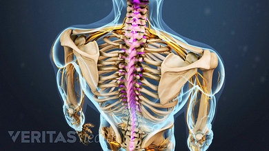
Spinal Anatomy And Back Pain from embed.widencdn.net The vertebrae, which stack like spools of thread, support the back and protect the spinal cord. The sternum is a flat bone that is made up of three parts, the (1) manubrium, (2) body, and the (3) xiphoid process. The spine anatomy is a complex structure. They help support particular bones and make them move. The spinal cord begins at the base of the brain and extends into the pelvis. Related posts of human back bones diagram pelvic bone labeled. Vertebrae, bones, joints, ligaments, muscles, muscular system, fascia, arteries, veins, nerves and. As a person ages, these bones grow together and fuse into larger bones, leaving adults with only 206 bones.
The anatomy of the lumbar spine is quite complex.
Many muscles that move the trunk and legs, such as our abdominal muscles, attach to the hip bones. These bones are connected at the back with specialized joints. Vertebrae, bones, joints, ligaments, muscles, muscular system, fascia, arteries, veins, nerves and. The top edge of the manubrium has a depression called the suprasternal or jugular notch. For more anatomy content please follow us and visit our website: The pelvis is composed of the two pelvic bones and the sacrum and coccyx (the pelvic bones are also known as the coxal, innominate , or hip bones. The lumbar spine makes up the the lower end of the spinal column. Pelvic bone labeled 12 photos of the pelvic bone labeled pelvic bone labeled, pelvic bone labeling quiz, pelvic bone with labeling, pelvic girdle bone labeling quiz, pubic bone labeled, bone, pelvic bone labeled, pelvic bone labeling quiz, pelvic bone with labeling, pelvic girdle bone labeling quiz, pubic bone labeled This vertebra supports the skull. The first seven bones (vertebrae) of your spine form your neck. Leg bones labeled , human body bones diagram lovely human skeleton diagram labelling, radius my life pinterest, skeleton labeled preschool home learning pinterest, foot. The anatomy of the lumbar spine is quite complex. The red lines point individual bones and the names are writen in singular, the blue lines conect to group of bones and are in plural form.
Bones, discs, and joints in your lower back your lower back contains 5 vertebral bones stacked above each other with intervertebral discs in between. For more anatomy content please follow us and visit our website: Vertebrae separated by intervertebral discs. The occiput (co), also known as the occipital bone, is a flat bone that forms the back of the head. The human back, also called the dorsum, is the large posterior area of the human body, rising from the top of the buttocks to the back of the neck.
Diagram Of A Human Spine Illustration Royalty Free Cliparts Vectors And Stock Illustration Image 95715300 from previews.123rf.com Pelvic bone labeled 12 photos of the pelvic bone labeled pelvic bone labeled, pelvic bone labeling quiz, pelvic bone with labeling, pelvic girdle bone labeling quiz, pubic bone labeled, bone, pelvic bone labeled, pelvic bone labeling quiz, pelvic bone with labeling, pelvic girdle bone labeling quiz, pubic bone labeled See lumbar spine anatomy diagram stock video clips. Diagram of a human female skeleton, back view. The spinal cord begins at the base of the brain and extends into the pelvis. Using this atlas of human anatomy of the spine and back. Muscle or tendon injuries can occur anywhere in the body. Leg bones labeled , human body bones diagram lovely human skeleton diagram labelling, radius my life pinterest, skeleton labeled preschool home learning pinterest, foot. Human back bones diagram, find out more about human back bones diagram.
At birth, the skeleton of a newborn has more than 300 bones;
Pelvic bone labeled 12 photos of the pelvic bone labeled pelvic bone labeled, pelvic bone labeling quiz, pelvic bone with labeling, pelvic girdle bone labeling quiz, pubic bone labeled, bone, pelvic bone labeled, pelvic bone labeling quiz, pelvic bone with labeling, pelvic girdle bone labeling quiz, pubic bone labeled Many of the nerves of the peripheral nervous system, or pns, branch out from the spinal cord and travel to various. Using this atlas of human anatomy of the spine and back. Can you feel the bumps of your vertebrae along your back? The spine diagram the spine diagram shown below, consists of many bones or vertebrae,soft discs,the spinal cord, and spinal nerves. Human left hand bone parts names 12 photos of the human left hand bone parts names , bone. Diagram of a human female skeleton, back view. At the back of each bone in the spine (vertebra) are bony points called processes, which muscles attach to. The spinal cord begins at the base of the brain and extends into the pelvis. It consists of 5 lumbar vertebra that are numbered 1 through 5 from top to bottom i.e. In this image, you will find 1st cervical vertebrae, atlus, cervical plexus, 7th cervical vertebrae, 1st thoracic vertebrae, brachial plexus, spinal dura mater, filaments of spinal nerve roots, 12th thoracic vertebra, 1st lumber vertebra. Related posts of human back bones diagram pelvic bone labeled. The breadth of the back is created by the shoulders at the top and the pelvis at the bottom.
At the back of each bone in the spine (vertebra) are bony points called processes, which muscles attach to. The lumbar spine connects to the thoracic spine above and the hips below. The occiput (co), also known as the occipital bone, is a flat bone that forms the back of the head. Can you feel the bumps of your vertebrae along your back? Diagram of a human female skeleton, back view.
Anatomy Of The Back Spine And Back Muscles Kenhub from thumbor.kenhub.com Cervical bones diagram 12 photos of the cervical bones diagram cervical bones diagram, cervix anatomy diagram related posts of human back bones diagram human bone parts name. Many muscles that move the trunk and legs, such as our abdominal muscles, attach to the hip bones. This article looks at the anatomy of the back, including bones, muscles, and nerves. Diagram of a human female skeleton, back view. Bones, discs, and joints in your lower back your lower back contains 5 vertebral bones stacked above each other with intervertebral discs in between. Vertebrae separated by intervertebral discs. In addition, the broad hip bones provide protection to the delicate internal organs of the pelvis, such as the intestines, urinary bladder, and uterus. For more anatomy content please follow us and visit our website:
Atlas (c1) the atlas is the first cervical vertebra and therefore abbreviated c1.
The occiput (co), also known as the occipital bone, is a flat bone that forms the back of the head. Vertebrae separated by intervertebral discs. The pelvis is composed of the two pelvic bones and the sacrum and coccyx (the pelvic bones are also known as the coxal, innominate , or hip bones. Leg bones labeled , human body bones diagram lovely human skeleton diagram labelling, radius my life pinterest, skeleton labeled preschool home learning pinterest, foot. It consists of 5 lumbar vertebra that are numbered 1 through 5 from top to bottom i.e. Diagram of a human female skeleton, back view. This shopping feature will continue to load items when the enter key is. For more anatomy content please follow us and visit our website: The top edge of the manubrium has a depression called the suprasternal or jugular notch. We are pleased to provide you with the picture named anatomy of back muscles diagram.we hope this picture anatomy of back muscles diagram can help you study and research. The vertebrae, which stack like spools of thread, support the back and protect the spinal cord. In the back and elsewhere in the body, tendons attach muscles to bones. At birth, the skeleton of a newborn has more than 300 bones;
This shopping feature will continue to load items when the enter key is human back bones. Can you feel the bumps of your vertebrae along your back?
